Wednesday, December 26, 2012

Skin care management in cancer patients: an evaluation of quality of life and tolerability

Skin care management in cancer patients: an evaluation of quality of life and tolerability

April 2011

Abstract

Keywords:  Skin care, Chemotherapy, Cancer, Dry skin, Radiation therapy, Skin irrations, HFSR

Purpose: The objective of this study is to evaluate quality of life (QoL) and tolerability of three articles specifically developed for cancer skin care management (skin moisturizer, face moisturizer, and face wash).

Methods: Participants were cancer patients (n = 99) receiving systemic anticancer therapies and/or radiotherapy at Northwestern University. Subjects were assessed at the initial visit for adverse skin reactions based on the National Cancer Institute’s Common Terminology Criteria for Adverse Events version 3.0 and completed the Skindex-16 questionnaire, a self-reported dermatology-specific QoL instrument. All subjects were provided with three test articles and were instructed to use each test article once daily for 4 weeks. At the 4-week follow-up (n = 77), the Skindex-16 was readministered, adverse skin reactions were assessed, and tolerability questionnaires were administered for each article used.


Results: Dry skin, hand–foot skin reaction (HFSR), and skin rash (dermatitis) decreased significantly from baseline to follow-up. Presence of nail changes, skin rash (desquamation), and acne/acneiform eruptions did not significantly change from baseline. Subjects had a significantly lower mean overall Skindex-16 score at 4-week follow-up when compared to baseline. Most patients rated their overall experience with each test article as good or very good (highest rating).


Conclusion: Skin care in cancer patients is suboptimal in part due to a lack of products and knowledge specific for this population. Our findings suggest that QoL improves with test article use, all of which were rated as good/very good for tolerability. Moreover, skin toxicity as manifested by dry skin, hand-foot skin reaction, and skin rash (dermatitis) were decreased with use of test articles within 4 weeks.

Full Text

Saturday, December 22, 2012

Complex Decongestive Physiotherapy Treats Skin Changes like Hyperkeratosis Caused by Lymphedema.


Complex Decongestive Physiotherapy Treats Skin Changes like Hyperkeratosis Caused by Lymphedema.


2012

Source

School of Physical Therapy and Rehabilitation, Abant Izzet Baysal University, 14280 Bolu, Turkey.

Abstract


Lymphedema is a chronic, progressive, and often debilitating condition. Primary lymphedema is a lymphatic malformation developing during the later stage of lymph angiogenesis. Secondary lymphedema is the result of obstruction or disruption of the lymphatic system, which can occur as a consequence of tumors, surgery, trauma, infection, inflammation, and radiation therapy. Here, we report a 64-year-old woman presenting with hyperkeratosis, a lymphedema due to metastatic uterus carcinoma. In this paper, we present the effects of complex decongestive physiotherapy on lymphedema and hyperkeratosis.

Friday, December 14, 2012

Wednesday, December 5, 2012

Safety and efficacy of personal care products containing colloidal oatmeal.


Safety and efficacy of personal care products containing colloidal oatmeal.


2012

Source

Johnson & Johnson Santé Beauté France, Issy les Moulineaux, France.

Abstract


BACKGROUND:

Colloidal oatmeal is a natural ingredient used in the formulation of a range of personal care products for relief of skin dryness and itchiness. It is also used as an adjunctive product in atopic dermatitis. The safety of personalcare products used on vulnerable skin is of particular importance and the risk of developing further skin irritations and/or allergies should be minimized.

METHODS:

In a series of studies, we tested the safety of personal care products containing oatmeal (creams, cleansers, lotions) by assessing their irritant/allergenic potential on repeat insult patch testing, in safety-in-use and ocular studies using subjects with nonsensitive and sensitive skin. We also tested the skin moisturizing and repair properties of an oatmeal-containing skin care product for dry skin.

RESULTS:

We found that oatmeal-containing personal care products had very low irritant potential as well as a very low allergenic sensitization potential. Low-level reactions were documented in 1.0% of subjects during the induction phase of repeat insult patch testing; one of 2291 subjects developed a persistent but doubtful low-level reaction involving edema during the challenge phase in repeat insult patch testing. No allergies were reported by 80 subjects after patch testing after in-use application. Sustained skin moisturizing was documented in subjects with dry skin that lasted up to 2 weeks after product discontinuation.

CONCLUSION:

Our results demonstrate that colloidal oatmeal is a safe and effective ingredient in 

Sunday, November 25, 2012

Common Skin Infections

Common Skin Infections


Nathaniel C. Cevasco

Kenneth J. Tomecki


Bacterial infections

Humans are natural hosts for many bacterial species that colonize the skin as normal flora.Staphylococcus aureus and Streptococcus pyogenes are infrequent resident flora, but they account for a wide variety of bacterial pyodermas. Predisposing factors to infection include minor trauma, preexisting skin disease, poor hygiene, and, rarely, impaired host immunity.

Impetigo

Definition and Etiology
Impetigo is a superficial skin infection usually caused by S. aureus and occasionally by S. pyogenes.

Prevalence and Risk Factors
Impetigo affects approximately 1% of children.

Pathophysiology and Natural History
S. aureus produces a number of cellular and extracellular products, including exotoxins and coagulase, that contribute to the pathogenicity of impetigo, especially when coupled with preexisting tissue injury. Impetigo commonly occurs on the face (especially around the nares) or extremities after trauma.

**Also included in this article:
Folliculitis, Furunculosis, Carbunculosis, Candidiasis, Ecthyma, Erysipelas, Cellulitis, Necrotizing Fasciitis, Dermatophytosis, Herpes Zoster, Tinea (Pityriasis) Versicolor, Viral infections**

Sunday, November 11, 2012

Clinical symptoms and therapy of necrotizing skin and soft tissue infections


Clinical symptoms and therapy of necrotizing skin and soft tissue infections


Nov 2012
[Article in German]

Source

Klinik für Chirurgie, Universitätsklinikum Schleswig-Holstein, Campus Lübeck, Ratzeburger Allee 160, 23538, Lübeck, Deutschland, peter.kujath@chirurgie.uni-luebeck.de.

Abstract


Skin and soft tissue infections are among the most common diseases requiring surgical treatment. The presentation of patients varies from folliculitis to severe necrotizing infections with a fatal outcome. The diagnosis of a necrotizing infection is often difficult. The correct diagnosis is often made after deterioration of the patient's condition in the rapid course of the disease. The early and correct diagnosis and immediate surgery are decisive for the prognosis. Treatment at a specialized intensive care unit and the administration of a broad spectrum antibiotic are pivotal for the survival of individual patients.

Skin and soft-tissue infections caused by Aeromonas species.


Skin and soft-tissue infections caused by Aeromonas species.


Nov 2012

Source

Department of Intensive Care Medicine, Chi Mei Medical Center, Liouying, Tainan, Taiwan.

Abstract


This study investigated the clinical characteristics of patients with skin and soft-tissue infections (SSTIs) due to Aeromonas species. Patients with SSTIs caused by Aeromonas species during the period from January 2009 to December 2011 were identified from a computerized database of a regional hospital in southern Taiwan. The medical records of these patients were retrospectively reviewed. A total of 129 patients with SSTIs due to Aeromonas species were identified. A. hydrophila (n = 77, 59.7 %) was the most common pathogen, followed by A. veronii biovar sobria (n = 22, 17.1 %), A. veronii biovar veronii (n = 20, 15.5 %), A. caviae (n = 9, 7.0 %), and A. schubertii (n = 1, 0.8 %). The most common isolates obtained from patients with polymicrobial infections were Klebsiella species (n = 33), followed by Enterococcus spp. (n = 24), Enterobacter spp. (n = 21), Escherichia coli (n = 17), Staphylococcus spp. (n = 17), Streptococcus spp. (n = 17), and Acinetobacter spp. (n = 15). Liver cirrhosis and concomitant bacteremia were more common among patients with monomicrobial Aeromonas SSTIs than among patients with polymicrobial SSTIs. Nine (7 %) patients required limb amputations. The in-hospital mortality rate was 1.6 %. In conclusion, Aeromonas species should be considered as important causative pathogens of SSTIs, and most infections are polymicrobial. In addition, the clinical presentation differs markedly between patients with monomicrobial and those with polymicrobial Aeromonas SSTIs.

Systemic antioxidants and skin health.


Systemic antioxidants and skin health.


Sept 2012

Abstract


Most dermatologists agree that antioxidants help fight free radical damage and can help maintain healthy skin. They do so by affecting intracellular signaling pathways involved in skin damage and protecting against photodamage, as well as preventing wrinkles and inflammation. In today's modern world of the rising nutraceutical industry, many people, in addition to applying topical skin care products, turn to supplementation of the nutrients missing in their diets by taking multivitamins or isolated, man-made nutraceuticals, in what is known as the Inside-Out approach to skin care. However, ingestion of large quantities of isolated, fragmented nutrients can be harmful and is a poor representation of the kind of nutrition that can be obtained from whole food sources. In this comprehensive review, it was found that few studies on oral antioxidants benefiting the skin have been done using whole foods, and that the vast majority of current research is focused on the study of compounds in isolation. However, the public stands to benefit greatly if more research were to be devoted toward the impact that physiologic doses of antioxidants (obtained from fruits, vegetables, and whole grains) can have on skin health, and on health in general.

Tuesday, November 6, 2012

Probiotics Boosts Skincare Regimen


Probiotics Boosts Skincare Regimen


Probiotics help maintain a healthy body -- but what about using probiotics in a regular skincare regimen? The latest article by Skincare-News.com Probiotics: Learn How Some Bacteria Can be Good for Your Skin explains how the beneficial bacteria in probiotics can help create and sustain a clear, healthy complexion for all skin types. From severe acne to preventing anti-aging, these valuable bacteria are a beautifying addition to any skincare regimen.

Using probiotics as part of a healthy skincare regimen is becoming more and more popular. What makes probiotics a heavenly find for skincare, and how can probiotics found in skincare products, supplements and foods treat skin conditions such as acne and eczema? Plus, for those concerned with the signs of aging, how do probiotics contribute to the effectiveness of anti-aging products? 

One of the greatest benefits of adding probiotics to a daily routine is its convenience and accessibility. Found in many forms, from staple foods like yogurt and pickles to anti-aging serums, there are various ways to integrate probiotics into a daily routine. What are the best probiotic-containing foods, and what are some tips for ensuring maximum benefits?

Skin care tips to combat the effects of cold weather


Skin care tips to combat the effects of cold weather


By Tiffany Bentley | The Express-Times 
on November 04, 2012 at 7:06 AM, updated November 04, 2012 at 7:11 AM

Colder weather can take its toll even on the healthiest skin. Laura Parker, esthetician and owner of The Skin Clinic in Easton, offers the following basic tips on how to make sure the season doesn't diminish your glow

  • Switch to a heavier lotion and cleanse the skin only once a day at night to prevent dryness. Over-cleansing the skin can actually strip the skin of necessary oils that keep it looking healthy.
  • Try to take shorter showers. Winter usually means we take hotter showers, which zaps the skin of moisture. Make sure to moisturize head to toe immediately after and towel dry. The skin will be able to absorb the moisturizer more effectively if it is still moist.
  • Don’t skip sunscreen. This is one of the biggest skin care mistakes people make during the winter. UVA and UVB rays are still out in the winter and you can even catch damage through your windshield driving to and from work. If you plan on spending a lot of time outside, such as skiing, make sure to wear an SPF of 30 or higher.
  • Exfoliate. One of the best ways to prepare skin for winter and repair damage caused by the summer is exfoliation. Alpha hydroxy acids (AHAs) are my favorite form of exfoliation because they dissolve dead skin without the irritation of a harsh scrub. AHAs are also water-loving. After you use them they actually help bind moisture to the skin. They also increase cellular turnover, improving skin tone and texture. Common AHAs are glycolic acid (derived from sugar), lactic acid (derived from milk) and mandelic acid (derived from almonds). The Skin Clinic offers AHA exfoliating pads that retail at $15.
  • Stay hydrated. Drink at least six to eight glasses of water a day. A healthy dose of water increases the body’s ability to remove toxins, which can lead to acne breakouts, dry skin and inflammation.
  • Monitor the heater. As the weather becomes cold, we tend to crank up the heat, which can throw your skin's balance out of whack. The combination of an indoor heater and drier weather can cause skin to become dehydrated and as a result, send oil production into overdrive. This increase in oil or sebum production and skin dehydration can cause pore-clogging buildup and lead to breakouts.
  • Treat yourself to a facial. Facials provide a professional level of exfoliation, balancing and deep cleansing the skin while addressing a multitude of skin conditions including acne, rosacea and prematurely aging skin. 

Thursday, November 1, 2012

Lymphomatoid papulosis

Lymphomatoid papulosis


Related Terms and Key Words: CD30+ lymphoma, eccrinotropic, granulomatous, lymphomatoid papulosis, methotrexate, lymphedema, papulonodular skin eruption, Primary cutaneous CD30 + lymphoproliferative disorder, Cutaneous T-cell lymphoma, Proliferative T-cell disorder, WHO/EORTC classification, chemokines, chemokine receptors
Definition:
A chronic skin disease that presents with characterists of malignant T-cell lymphoma. However, it is important to remember that lymphotoid papulosis (LyP) is not classified as a true lymphoma. Also, it does not spread and is not fatal. It is therefore described as histologically malignant, but clinically benign. (1)
It is classified as a lymphoproliferative disorder. These account for about 25% of cutaneous T-cell lymphomas. The condition occurs equally between men and women and usually occurs in the fifth decade. Clinically, black people appear to have the disease much less frequently then other races.
The term lymphomatoid papulosis originally was used by Macaulay[1] in 1968 to describe “a self-healing rhythmical paradoxical eruption, histologically malignant but clinically benign.” Due to the typical waxing and waning clinical course, lymphomatoid papulosis was previously considered a pseuodolymphomatous inflammatory process. However, the classification system for cutaneous lymphomas has evolved rapidly, and, during consensus meetings in 2003-2004, the World Health Organization—European Organization for Research and Treatment of Cancer (WHO-EORTC) classification grouped lymphomatoid papulosis among the indolent cutaneous T-cell lymphomas. (2)
LyP is divided into three subtypes, they include:
Type A - characterized by large CD30 atypical cells intermingled with a prominent inflammatory infiltrate. The large tumor cells have polymorphic convoluted nuclei with a minimum of 1 prominent nucleolus and resemble Reed-Sternberg cells when binucleate, as is seen in HD. Type A lymphomatoid papulosis is the most common histologic variant and accounts for 75% of all lymphomatoid papulosis specimens.
Type B is characterized by smaller (8-15 µm) atypical cells with hyperchromatic cerebriform nuclei resembling the atypical lymphocytes in MF. CD30+ large cells are rare, but epidermotropism is more common in this variant. There is some concern that Type B lymphomatoid papulosis may be better classified as a papular variant of MF.
Type C (diffuse large cell type) is characterized by sheets of CD30+ anaplastic large cells indistinguishable from ALCL, with the exception of the minimal subcutaneous invasion. These lesions resolve spontaneously and are therefore classified as lymphomatoid papulosis; however, some authorities view this histologic variant as borderline ALCL or, perhaps, pcALCL. (2)
Uncommonly, patients may have more than one histologic subtype of lymphomatoid papulosis or other recently described associated histologic patterns.
Risk Factors:
Unknown at the present time and there is debate on whether or not it may be genetically caused. Several genetic defect have been identified in LyP lesions, but the specific cause has not been identified.
A few investigators have also discovered viruslike particles in lymphomatoid papulosis lesions examined under electron microscopy.
Signs and Symptoms:
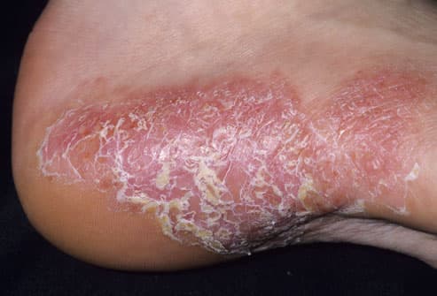
Lymphomatoid papulosis appears as recurrent small, raise skin lesions. The color ranges from red to brown in color. The most commonly occur on the trunk, arms, and legs in crops but may also occur on the palms, soles, face, and scalp. Very rarely, LyP lesions may be present in the mouth and throat. The lesions heal spontaneously within several weeks and leave a small, hypopigmented scar.
Diagnosis:
Done by skin biopsy. This is critical as the lesions may appear as the same for numerous other skin nodular growths.
Biopsies are also critical to rule out mycoses fungoides, cutaneous anaplastic large cell lymphoma, cutaneous Hodgkin’s disease, cutaneous leukemia, scabies, insect bites, and drug reactions.
If after biopsy the result come back other then lymphotoid papulosis, then the treating physician will order other types of tests to establish a correct diagnosis.
Differential Diagnosis
Cutaneous CD30+ (Ki-1) Anaplastic Large-Cell Lymphoma, Cutaneous T-Cell Lymphoma, Folliculitis, Insect Bites, Langerhans Cell Histiocytosis, Leukemia Cutis, Lymphocytoma Cutis, Milia, Miliaria, Scabies
Prognosis:
The disease itself is not fatal. However, 10 to 20% of patients will also develop an associated systemic lymphoma, typically anaplastic large cell, Hodgkinís disease, or mycoses fungoides.
If any type of secondary malignancy develops, then that would alter the outlook, depending on the type of malignancy.
Treatment:
Patients may choose not to treat the lesions and they usually heal spontaneously over 1-2 months.
Historically, treatment for the skin lesions themselves has been corticosteroid creams or ointment. Another option that can lead to faster healing is with low dose methotrexate. This is a type of chemotherapy that inhibits cell division. The one weak point in this treatment is that the lesions treated with methotrexate will generally return several weeks after therapy.
Another strong, aggressive treatment is oral psoralen plus ultraviolet light, so-called PUVA therapy.
Other treatments might include: carmustine, topical nitrogen mustard, topical MTX, topical imiquimod cream, intralesional interferon, low-dose cyclophosphamide, chlorambucil, medium-dose UVA-1 therapy, excimer laser therapy, photodynamic therapy, and dapsone help disease suppression.
Mortality/Morbidity
Lymphomatoid papulosis has a chronic, indolent course in most patients.
However, associated lymphomas may arise with LyP. These include immunoblastic lymphoma, lethal midline granuloma (currently considered as natural killer cell lymphoma in many patients), and systemic lymphocytic lymphoma. In most patients, the malignancy develops many years after the diagnosis of lymphomatoid papulosis.
(1) Lymphomatoid Papulosis - Know Cancer

Lymphomatoid Papulosis
see also:


Friday, October 19, 2012

Papillomatosis


Papillomatosis


These are small usually round nodules that seem to literally pop out of the skin on late stage lymphedema patients.  Normally, they are skin colored, can grow singularly or in clusters.  For the population at large, these are almost always benign growths that really require no followup or special care.

However, in lymphedema patients these can become angiosarcoma and thus need to be promptly removed with the nodules sent to pathology lab for examination.  

I have had many removed over the years and in fact just recently had well over twenty removed from the knee areas of both legs.  The surgeon "snips" the stem that these grow on and removes them.  For the most part, they can be removed on an outpatient basis.  There are no stitches involved either and after healing there is practically no scar either.

The dressing is simple.  I used an antibiotic ointment covered with a smooth bandage to go directly over the wound, then a layer of gauze.  Since my skin will not tolerate any type of tape, I keep it in place using my short stretch bandage on the leg.  Don't put gauze directly onto the wound as it will get stuck in the seepage of the wound and can cause damage to the wounds healing processes.

I would also recommend an antibiotic to be taken as a preventative measure to help insure there is no infection or cellulitis due to the immunocompromised state of a lymphedematous limb.  

Pat

----------------------


Abstracts:


Stasis papillomatosis. Clinical features, etiopathogenesis and radiological findings.


1993

Source

University Department of Dermatology, St. Josef Hospital, Bochum, Germany.

Abstract 


Stasis papillomatosis can be divided into localized and widespread forms. Ten patients with this disease are discussed with emphasis on their clinical and lymphoradiological findings. Using isotope lymphography we were able to find an overall lymphostasis in only four patients. In all patients, local lymphatic disturbances were detected by means of indirect lymphography with intradermal lymph cysts as the most specific sign. Local dermal lymphostasis seems to be the common final pathogenesis in spite of different etiologies (ie, primary lymphedema, chronic venous insufficiency, trauma recurrent erysipelas, and local lymphangiodysplasia of unknown origin). A maximum variant was seen, following en-bloc resection of subcutaneous tissue in a patient suffering from congenital lymphedema.
PubMed


Ileostomy-Associated Chronic Papillomatous Dermatitis Showing Nevus Sebaceous-Like Hyperplasia, HPV 16 Infection, and Lymphedema: A Case Report and Literature Review of Ostomy-Associated Reactive Epidermal Hyperplasias.


---------------------------------------


Diagnostic Images:


Yahoo

DermIS

Thursday, September 20, 2012

Skin Care - The Big Four Amino Acids

Skin Care - The Big Four Amino Acids

Proline P 


Proline shares many properties with the aliphatic group.

Proline is formally NOT an amino acid, but an imino acid. Nonetheless, it is called an amino acid. The primary amine on the α carbon of glutamate semialdehyde forms a Schiff base with the aldehyde which is then reduced, yielding proline.

When proline is in a peptide bond, it does not have a hydrogen on the α amino group, so it cannot donate a hydrogen bond to stabilize an α helix or a β sheet. It is often said, inaccurately, that proline cannot exist in an α helix. When proline is found in an α helix, the helix will have a slight bend due to the lack of the hydrogen bond.

Proline is often found at the end of α helix or in turns or loops. Unlike other amino acids which exist almost exclusively in the trans- form in polypeptides, proline can exist in the cis-configuration in peptides. The cis and trans forms are nearly isoenergetic. The cis/transisomerization can play an important role in the folding of proteins and will be discussed more in that context.


Glycine

Glycine is one of the non-essential amino acids and is used to help create muscle tissue and convert glucose into energy. It is also essential to maintaining healthy central nervous and digestive systems, and has recently been shown to provide protection via antioxidants from some types of cancer. 

Glycine is used in the body to help construct normal DNA and RNA strands—the genetic material needed for proper cellular function and formation. It helps prevent the breakdown of muscle by boosting the body’s levels of creatine, a compound that helps build muscle mass. High concentrations of glycine are found not only in the muscles, but in the skin and other connective tissues as well. Almost 1/3 of collagen, which keeps the skin and connective tissue firm and flexible, is composed of glycine. (High amounts of Glycine are also found in gelatin, which is a form of denatured collagen). Without glycine the body would not be able to repair damaged tissues; the skin would become slack as it succumbed to UV rays, oxidation, and free radical damage, and wounds would never heal. 

Glycine is considered a glucogenic amino acid, which means it helps supply the body with glucose needed for energy. It helps regulate blood sugar levels, and thus glycine supplementation may be useful for treating symptoms characterized by low energy and fatigue, such as hypoglycemia, anemia, and Chronic Fatigue Syndrome (CFS). 

Glycine is essential for a healthy, normally functioning digestive system. It helps regulate the synthesis of the bile acid used to digest fats, and is included in many commercial gastric antacid agents. 

Glycine is necessary for central nervous system function. Research has shown that this amino acid can help inhibit the neurotransmitters that cause seizure activity, hyperactivity, and manic (bipolar) depression. Glycine can also be converted to another neurotransmitter, serine, as needed, and may be beneficial in the management of schizophrenia. In one study, twenty-two schizophrenic patients, who did not initially respond to traditional treatments, added glycine to their ongoing antipsychotic medication and found that it significantly reduced their symptoms. Glycine intake among the participants ranged from 40 to 90 grams daily (0.8 grams per kilogram of body weight). More research concerning the effects of glycine on schizophrenia is underway. Studies have shown that glycine also helps improve memory retrieval loss in those that suffer from a wide variety of sleep-depriving conditions, including schizophrenia, Parkinson’s disease, Huntington’s disease, jet lag, and overwork. 

Results from preliminary studies of glycine as a potential treatment for cancer have been promising, and suggest that it may help prevent the development of cancerous tumors and melanoma. In laboratory mice, dietary glycine prevented tumor growth by inhibiting angiogenisis, the process by which tumors develop their own blood supply. Glycine also seems to play a role in keeping the prostate healthy. In one study, glycine was shown to help reduce the symptoms of prostatic hyperplasia in men. 

High-protein foods, such as fish, meat, beans, milk, and cheese, are the best dietary sources of glycine. Glycine is also available in capsule and powder forms, and as part of many combination amino acid supplements. There have been no toxic effects associated with glycine, although some people have reported that taking this supplement causes stomach upset. 

Individuals with kidney or liver disease should not consume glycine without consulting their doctor. Taking any one amino acid supplement can cause a disruption of the citric acid or Krebs cycle, and cause a build-up of nitrogen or ammonia in the body, which makes the liver and kidneys work harder to remove waste. Anyone taking antispastic drugs should consult a physician before supplementing with glycine, since it theoretically could increase the effects of these medications. 


Leucine

Leucine, an essential amino acid, is one of the three amino acid with a branched hydrocarbon side chain. It has one additional methylene group in its side chain compared with valine.


Leucine (abbreviated as Leu or L)[2] is a branched-chain α-amino acid with the chemical formula HO2CCH(NH2)CH2CH(CH3)2. Leucine is classified as a hydrophobic amino acid due to its aliphatic isobutyl side chain. It is encoded by six codons (UUA, UUG, CUU, CUC, CUA, and CUG) and is a major component of the subunits in ferritinastacin and other 'buffer' proteins. Leucine is an essential amino acid, meaning that the human body cannot synthesize it, and it therefore must be ingested.

Info Page: Wikipedia

Lysine

Overview:

Lysine, or L-lysine, is an essential amino acid. That means it is necessary for human health, but the body can't manufacture it. You have to get lysine from food or supplements. Amino acids like lysine are the building blocks of protein. Lysine is important for proper growth, and it plays an essential role in the production of carnitine, a nutrient responsible for converting fatty acids into energy and helping to lower cholesterol. Lysine appears to help the body absorb calcium, and it plays an important role in the formation of collagen, a substance important for bones and connective tissues including skin, tendon, and cartilage.
Most people get enough lysine in their diet, although athletes, vegans who don't eat beans, as well as burn patients may need more. Not enough lysine can cause fatigue, nausea, dizziness, loss of appetite, agitation, bloodshot eyes, slow growth, anemia, and reproductive disorders. For vegans, legumes (beans, peas, and lentils) are the best sources of lysine.

Dietary Sources:

Foods rich in protein are good sources of lysine. That includes meat (specifically red meat, pork, and poultry), cheese (particularly parmesan), certain fish (such as cod and sardines), nuts, eggs, soybeans (particularly tofu, isolated soy protein, and defatted soybean flour), spirulina, and fenugreek seed. Brewer's yeast, beans and other legumes, and dairy products also contain lysine. Many nuts also contain lysine along with arginine (lysine counteracts some of the effects of arginine). So if someone is trying to eat a diet rich in lysine to prevent HSV outbreaks, nuts would be a good choice.

Full text Page:  University of Maryland Medical Center